Articles by "gynecology"
Showing posts with label gynecology. Show all posts

Polyhydramnios And Oligohydramnios


Polyhydramnios


Polyhydramnios
Polyhydramnios

Polyhydramnios

Definition:


Is an excessive amount of amniotic fluid more than 2000 ml.

The exact cause is unknown, it is associated with:


- Maternal disease as DM, Renal disease.

- Multiple pregnancies.

- Fetal abnormalities that affect the swallowing mechanism.

- The condition leads to preterm delivery, malpresentation, and cord prolapse and abruptio placenta, thus increasing maternal mortality.

Clinical manifestations:

- Excessive uterine enlargement, fundal height increases out of proportion to gestational age.

- Difficulty in breathing.

- Difficulty finding a comfortable sleeping position.

- Difficult to hear FHR and palpate fetus.

- Pain in the abdomen, back and thighs due to increasing pressure.

- Difficult ambulating.

- Varicosities.

- Nausea and vomiting.

Management:

- Hospitalization, if the mother is dyspneic or in pain.

- Transabdominal or vaginal amniocentesis with aid of sonography and careful monitoring of vital signs. remove the fluid slowly to avoid abruptio placenta.

- Offer support by explaining procedures.

- Encourage the women to rest on the side in a semi-recumbent position to increase blood flow to uterus and fetus and to relieve symptoms.

- Watch carefully for signs of the abruption placenta, abnormal presentation and post-partum haemorrhage.


Oligohydramnios 


Oligohydramnios
Oligohydramnios

Oligohydramnios

Definition:

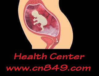
Is a small amount of amniotic fluid less than 400 ml.

It is associated with:

a- Fetal renal agenesis.

b- Post maturity.

Clinical manifestations:

- Small uterine size.

- Labour may be premature.

- Uterine contraction may be ineffectual and labour prolonged.

- Fetal hypoxia may occur because of cord compression.

Management:

- Monitor fetal status carefully during pregnancy and labour.

- Monitor the woman for labour complications.


READ MORE:


Pneumonia

Pleurisy and Peural Effusion

Laryngitis And Laryngeal Cancer

Preterm Labor


Is defined as rhythmic uterine contractions that produce cervical changes prior to completion of 37 weeks of gestation.


Preterm Labor
Preterm Labor

Aetiology:

1- Demographics:

- Upper and lower extremes of age.

- Lower socioeconomic status.

- Inadequate prenatal care.

- Race ( increase in blacks).

2- lifestyle and employment.

- Smoking and drug abuse.

- Prolonged periods of standing.

- Fatigue and long hours of work.

- Heavy work and lifting.

3- Reproductive history:

- Previous preterm delivery.

- Incompetent cervix.

- Spontaneous or induced abortion.

4- Uterine anomalies e.g. leiomyomata.

5- Weight again: low weight or low weight gain may increase risk.

6- Anemia: probably due to other risk factors.

7- Uterine size and placental abnormalities:

- Multiple gestations.

- Placenta previa.

- Polyhydramnios.

- Abruptio placenta.

8- Premature rupture of membranes.

9- Vaginal bleeding.

10- Surgery: abdominal procedures.

11- Infection: UTI, pneumonia, malaria, typhoid fever, syphilis, gonorrhoea, amniotic fluid infection, vaginitis.

12- Other associations:

- Fetal gender ( male fetuses have a shorter gestation period).

- low magnesium level.

Assessment:

- Cervical dilation.

- Membranes: ruptured or not.

- Presences of severe preeclampsia and haemorrhage.

- Ultrasonography: to determine fetal gestational age, condition and weight.

Management and intervention:

- Special prenatal care for high-risk women.

a- Frequent visits for weeks 22 to 32.

b- Urine culture at 24 weeks.

c- Vaginal examination for pH.

d- Education on nutrition and preterm labour.

Signs and symptoms reinforced :

- Increase or change in vaginal discharge.

- Uterine contraction.

- Vaginal bleeding or leaking fluid.

Bed rest and hydration: increase uterine blood flow.

Continuous monitoring

Fetal maturation therapy: glucocorticoid therapy.


READ MORE:


Hydatidiform ( Vesicular ) mole

Hypotonic And Hypertonic Uterine Contraction

Contracted Pelvis and Cephalopelvic Disproportion


Hydatidiform Mole ( Vesicular )


Hydatidiform Mole
Hydatidiform Mole


It is a developmental anomaly of the placenta and the trophoplast in which the fertilized ovum deteriorates and the chorionic villi convert into a mass of clear grape-like vesicles.

It is one of the most common lesions anteceding choriocarcinoma, a malignant tumour of the trophoblast with a tendency toward rapid and widespread metastasis.

Aetiology:

The cause is unknown, but factors contributed are:
- Maternal age: below 20 years and above 45 years.
- Genetic factors.
- High parity and malnutrition.

Clinical classification:

Vesicular mole is classified into complete and partial moles according to the presence or absence of a fetus or embryo and its location or dissemination.

Clinical manifestation:

1- Bleeding: the most common sign and vary from spotting to profuse, continuous or intermittent red or brownish bloody discharge, about the 12th week of gestation, may also pass villi.

2- Enlargement of the uterus is out of proportion to what it normally is at a specific time in pregnancy.

3- Signs of Preeclampsia or Eclampsia earlier than 20 weeks of gestation.

4- Hyperemesis Gravidarum experienced by 30 % of women with this condition.

5- Pallor and dyspnea anaemia.

6- hCG titer is markedly increased beyond the 90th day of gestation when normally expected to drop.

7- Anxiety and tremors thyroid dysfunction due to high hCG.

8- Uterine discomfort due to overstretching.

9- Absent fetal heart tone.

10- Absent fetal parts ( except in partial mole ) found on ultrasound Or X-ray.

Diagnostic evaluation:

- Ultrasound is a diagnostic method of choice.

- A patient often presents with vaginal bleeding, uterine enlargement in absence of fetal heart tone.

- CBC: Hb, HCT and RBCs are decreased.

- Blood chemistries: renal, liver and thyroid function test.

- Chest x-ray.

- hCG titers are elevated up to 1 to 2 million IU in 24 hours.

Management:

1- Suction curettage has low complications rate with uterine size < 16 weeks.
Excessive uterine enlargement may be predisposed to pulmonary complications as preeclampsia and fluid overload.

2- Primary Hysterectomy:

- Patients who have completed childbearing and desire sterilization are good candidates.

- Reduces malignant sequelae from 20 % to 5 %.

3- Prophylactic chemotherapy:

- May reduce malignant sequelae in high-risk patients.
- Not routinely recommended in cases of the uncomplicated mole.

4- Blood transfusion: to correct anaemia and replace blood loss.

Follow up: follow up supervision at least for 1 year includes the following :

1- Hcg measurement as:

Once weekly until titers are negative for 3 consecutive weeks, then:

Once monthly for 6 months, then every 2 month for 6 months and every 6 months.

2- Chest x-ray to and detect metastases are done every month.

3- start contraceptive is the best choice.


READ MORE:


Episiotomy

Hypotonic And Hypertonic Uterine Contraction

Contracted Pelvis and Cephalopelvic Disproportion


Hypotonic And Hypertonic Uterine Contraction


Hypotonic uterine contraction ( inertia ):

Hypotonic labour is defined as less than 3 contractions of mild to moderate intensity occurring in a 10 minute period during the active phase of labour.

Cervical dilatation and descent of the fetus slow greatly or stop.

Aetiology:

- Such labour occurs when uterine fibres are overstretched from large baby, twins, hydramnios or multiparty.

- May also caused by administration of sedation or narcotics.

- Bowel or bladder distention.

Potential maternal effects:

- Exhaustion.

- Infection.

- Postpartum haemorrhage.

- Stress and psychological trauma.

Potential fetal effects:

- Fetal sepsis ( infection).

- Fetal and neonatal death.

Medical management:

Oxytocic stimulation of labour or prostaglandin stimulation.

Hypertonic uterine contraction:

- Occurs in the latent phase of labour, with an increase in the frequency of contractions and a decrease in their intensity.

- Contractions are extremely painful because of uterine muscle cell anoxia but are ineffective in dilating and effacing the cervix, which leads to maternal exhaustion.

- Contraction may interfere with uteroplacental exchange and lead to fetal distress and even death.

- Contractions may be uncoordinated and involve only portions of the uterus.

- Usually occurs before 4 cm dilation, cause not yet known, may be related to fear tension.

Potential maternal effects:

- Loss of control related to the intensity of pain and lack of progress.
- Exhaustion.

- Dehydration.

Potential fetal effect:

Fetal asphyxia with meconium aspiration death.

Medical management:

Analgesic (morphine, meperidine ) if membranes not ruptured fetopelvic disproportion isn’t present.


READ MORE:


Episiotomy

Breastfeeding

Contracted Pelvis and Cephalopelvic Disproportion


Contracted Pelvis and Cephalopelvic Disproportion


Contracted Pelvis and Cephalopelvic Disproportion


Contracted Pelvis:

Is one in which the bony funnel of women pelvis is too narrow at some point for the fetus to pass through.

The term used with pelvic diameter 1 cm or more less than normal except transverse diameter 2 cm, this may involve one or more diameters.

Causes:

- Growth retardation.

- Chronic disease e.g. T.B.

- Sever disease e.g. Anemia.

- Bone disease e.g. Rickets.

- Before pregnancy, pelvic measurement can be identified using clinical and x-ray pelvimetry.

- During pregnancy, sonography is used to measure the fetal head in relation to the pelvis.

- When measurements are minimal in one or more places, they are described as marginal pelvis if the infant has a moderately small head is in a normal position and contractions are forceful.

- When the measurement is marginal, the physician may decide to allow a trial labour for a few hours, vaginal delivery may be accomplished and the women are spared major surgery.

- If it is little or no progress in baby's descent cesarean birth is performed.

- The couple needs support to cope with the stress of complicated labour and participate in the decision regarding cesarean birth.


Cephalopelvic Disproportion:

Is fetal head to maternal pelvis discrepancy.

Fetopelvic disproportion term used with other than cephalic presentation.

When CPD is great, it is impossible for the fetus to pass.

CPD is suspected when labour is prolonged, cervical dilation and effacement are slow and engagement of the presenting part is delayed.

Contractions are monitored as well as the fetal heart rate.

Trial labour is allowed to continue only as long as dilation and decent progress.

If there is no progress, a cesarean birth is performed.

Maternal complication includes PROM, uterine rupture and necrosis of maternal soft tissue from the pressure of the fetal head.

Fetal complication includes cord prolapse, extreme moulding of the skull with possible fracture and intracranial haemorrhage.


READ MORE:


Episiotomy

Breastfeeding

Acute Glomerulonephritis


Episiotomy


Episiotomy
Episiotomy


Is an incision made in the perineum to enlarge the vaginal outlet? it serves the following purposes :

- Prevent tearing of the perineum, substitute a straight surgical incision for the laceration that may otherwise occur.

- Facilitate repair of laceration and promote healing.

- Minimize prolonged and severe stretching of the muscles supporting the bladder or rectum which may later lead to stress incontinence or vaginal prolapse.

- Shorten the second stage, which may be important for maternal reasons as, PIH or fetal reasons for persistent bradycardia.

- Enlarges the vagina in case manipulation is needed to deliver an infant for example in breech presentation or for application of forceps.

Types of Episiotomies:

The type of episiotomy is designated by site and direction of the incision.

1- Median:

- Is one most commonly employed?

- It is effective, easily repaired and generally the least painful.

- The incision is made in the middle of the perineum and directed toward the rectum.

- Is believed to heal with few complications, more comfortable for the women.

- If a long and large incision is needed during delivery, it may necessitate incision into the anal sphincter.

2- Mediolateral:

- The incision is made laterally in the perineum.

- This method avoids the anal sphincter if enlargement is nodded.

- The blood loss is greater, the repair is more difficult.


Assessment:

- The episiotomy site is inspected every 15 minutes during the first hour after delivery, then on a daily basis.

- The site is assessed of tenderness, redness, swelling and evidence hematoma.

Note :
Because suture used to repair episiotomy are of absorbable material they don’t need to be removed and no dressing is applied.


READ MORE:


Diarrhea

Breastfeeding

Acute Glomerulonephritis


Placenta Previa


Placenta Previa
Placenta Previa


Is the development of the placenta in the lower uterine segment, partially or completely covering the internal cervical os.

Classifications:

Traditionally categorized into 3 types:

1- Complete, total or central Previa: internal os entirely covered.

2- Partial placenta previa: internal os partially covered.

3- Marginal placenta previa: placenta reaches the edge of the internal os.

Aetiology:

- Specific causes are unknown.

Implantation may be affected by:

1. Abnormality of endometrial vascularity.

2. Delayed ovulation.

3. Prior endometrial trauma.

4. Multiple pregnancies.

5. Previous uterine surgery.

Clinical manifestations:

- Usually presents as painless vaginal bleeding in the 3rd trimester but can occur as early as 20 weeks of gestation bleeding occurs without warning in the absence of trauma.

- Blood loss from the first bleeding is rarely fatal, in each subsequent episode bleeding is heavier.

- Placenta previa may not cause bleeding until labour begins, or complete dilatation has occurred.

- Bleeding occurs earlier and is more profuse with total placenta previa.

Diagnostic evaluation:

- Painless vaginal bleeding is placenta previa until proven otherwise.

- Ultrasound is the diagnostic technique of choice.

- Definitive diagnostic by direct palpation of the placenta is not recommended.

Vaginal examination " double setup procedure ":

- Under sterile technique by the physician after diagnosis by ultrasound.

- Inconclusive ultrasound or only low –lying placenta noted.

Note: the term low-lying implantation is used when the placenta situated in the lower uterine segment but away from the os.

- Sterile speculum examination performed first.

- It is attempted only if the physician and the women are prepared for cesarean delivery in the operating room.

- If greater than 3cm dilated and no placenta covering os, then perform amniotomy ( by the physician).


READ MORE:


Bacterial Meningitis

Multiple Pregnancies


Abruptio Placentae


Abruptio Placentae
Abruptio Placentae


Premature separation of the normally implanted Placentae. separation occurs in the area of decidua basalis, most often in the 3rd trimester, but can happen any time after 20 weeks.

Incidence:

Complicates approximately 1% of pregnancies.

Is a serious disorder, accounts for about 15 % of all perinatal mortality " most common cause of intrapartum fetal death ".

Permanent neurologic impairment in 14% of surviving infants.

Classification:

The 3 types of Abruptio Placentae are:

1- Covert: Placentae separates in the centre and the bleeding is concealed.

2- Overt: blood pass from under the Placentae causing vaginal bleeding.

3- Placentae prolapse: total separation of Placentae with massive bleeding.

Aetiology:

Maternal hypertension.

PIH.

Cocaine-induced.

Maternal smoking.

Short umbilical cord.

Uterine anomalies.

Poor nutrition.

Physical work.

Trauma.

Amniotomy in patients with polyhydramnios.


Recurrent Risk:

- Tenfold increase in second pregnancy overpopulation risk.

- With 2 previous abruptions 25% chance of the third abruption.

Clinical manifestation:

- Vaginal bleeding (80% of the patient), blood remains concealed (20% of patients).

- Sudden onset of severe continuous abdominal pain and/or low back pain.

- Uterine contraction with a rigid, tender and irritable uterus.

- Amniotic fluid colour may be dark red.

- If bleeding is severe, hypofibrinogenemia may develop ( consumptive coagulopathy).

Fetal activity may be increased, because of fatal hypoxia . with severe complete abruption fetal heart tones may not be heard.

Complication: " accompany moderate to severe abruption "

1- hypovolemic shock.

- Pituitary necrosis ( Sheehan Syndrome).

- Renal failure.

Aetiology: is unclear, probably from reduced renal perfusion.

2- Fetal hypoxia or anoxia with possible fetal death.

3- Consumptive coagulopathy hypofibrinogenemia DIC.

4- Covelair uterus: bleeding into the myometrium resulting in broad like the rigidity of the uterus.

5- Hepatitis post blood or fibrinogen transfusion.

Management:

1- maternal urine greater than 30ml/ hr.

2- HCT greater than 30 %.

3- induction of labour.

4- cesarean section: if

- Continued bleeding.

- Fetal distress.

- Maybe dangerous is the setting of coagulation defect.


READ MORE:

Multiple Pregnancies

Bacterial Meningitis

Croup and Epiglottitis


Multiple Pregnancies


Twin
Multiple Pregnancies


Twin gestation:

Types of twining:

1- monozygotic ( identical): are identical because they develop from fertilization of one ovum " the same sex " occurs at random in about 3 to 4 / 1000.

2- dizygotic ( fraternal): twining occurs more frequently in some families " heredity is important on mother side".

- Occurs in response to greater levels of FSH.

- Increased in women greater than 35 years of age and in obese women.

- More common among Africans.

- Maybe different sexes.

- Always have 2 chorines, 2 amnions.

- The result of fertilization of 2 separate ova.

Note:
Conjoined twins with shared organs occur as a monozygotic twin through a division of the fertilized ovum after the 13th days post conception.

- A greater increase in blood volume, pulse, cardiac output and weight gain.

- Increased rate of preterm labour, hypertension, abruption, anaemia, hydramnios, UTI, cesarean section and postpartum haemorrhage.


Infant complications:


Prematurity – the average age of delivery, 37 weeks.

Discordance: defined as a difference of greater than 20% to 25% in weight.

Aetiology:

- The difference in the placental surface area.

-Twin to twin transfusion syndrome.

- Donor twin small, pale, anaemia.

- Recipient twin large, plethoric, polycythemia, hyperbilirubinemia.

- Fetal anomalies occur more often in multiple pregnancies.

Clinical manifestations:

1- Uterus larger than expected for the length of gestation.

2- Two fetal heart tones can be counted simultaneously.

3- Abdominal palpation yields many small parts by 6-7 month.

4- U/S usually used to confirm the diagnosis.

5- Oversized uterus and increased abdominal pressure often lead to:

- Digestion difficulty.

- Constipation.

- Dyspnea.

- A backache.

- Haemorrhoids and other varicosities.

Triplets:

- Average weight gains 45 to 50 pounds.

- Usual spontaneous time for delivery is 32-34 weeks.

- Average weight 1800 -1900 grams.

- Most delivered by cesarean section.

Quadruplets or more:

- Average weight gains 50 to 55 pounds.

- Average gestational age 30 to 31 weeks.

- Average weight 1200 – 1500 grams.

- The multifetal reduction has been shown to improve perinatal survival rate.


READ MORE:


Spina Bifida

Otitis Media

Croup and Epiglottitis


Ectopic Pregnancy


Ectopic Pregnancy
Ectopic Pregnancy


Is any gestation located outside the uterine cavity? when a fertilized ovum implants any place other than the endometrium of the uterus, the pregnancy called ectopic or extrauterine.

Most common cause of maternal death in the first half of pregnancy but, mortality is decreasing.

Factors responsible for increasing incidence ( diagnosis ) of ectopic pregnancy:

- Pelvic inflammatory disease.

- Improved diagnostic method.

- Sensitivity and specific hCG assays.

- High-resolution ultrasound.

- Diagnostic lumbar scoby.

- Increase awareness.

Aetiology:

Ectopic implantation may be fortuitous or result of tubal abnormality, which obstructs or delays the passage of the fertilized ovum as :

- Previous tubal surgery.

- Preceding pelvic inflammatory disease.

- IUD.

- Salpingitis.

- Congenital anomalies of the tube.

- Migration of the ovum across the pelvic cavity to the fallopian tube on the opposite side.

Frequency and implantation site:

1- tubal pregnancy: most frequent location, 86% in the distal half.

- Ampullary: the most common.

- Isthmus: early rupture.

- Interstitial: rare but very dangerous because it ends in rupture uterus and haemorrhage.

- Infundibular.

2- Abdominal pregnancy: mortality rate much higher.

3- Ovarian pregnancy: pregnancy attached to the uterus by utero-ovarian vasculature. Oophorectomy may be indicated.

4- Cervical pregnancy: implantation within the endocervical canal, very rare.

Clinical manifestation:

Very with the site of implantation and usually occur after tubal rupture.

Early signs and symptoms:

- Menstrual irregularities ( irregular vaginal bleeding).

- Symptoms of early pregnancy.

- Dull pain on the affected side.

Signs and symptoms of the tubal rupture:

- Pain: sudden, severe, and unilateral, generalized and radiated to the shoulder and neck due to phrenic nerve stimulation.

- Vaginal bleeding: dark brown and scanty, about 25 % of cases without vaginal bleeding.

Nausea, vomiting, fainting ( signs of internal blood loss ).

- Signs of shock.

- Normal or low temperature.

- Ruptured tubal pregnancy from salpingitis.

- Tenderness over abdomen upon palpation .

- Pelvic mass posterior or lateral to uterus .

- Cervical pain during vaginal examination .

- Distension of posterior fornix with blood in the cul-de-sac.

Diagnostic evaluation:

- Medical history.

- Physical examination.

- HCG assay: serum hCG usually lower than normal.

- Laparoscopy: often the diagnosis made by direct visualization can be performed too early.

Culdocentesis:

1- Needle puncture into posterior cul-de-sac.

2-  Negative culdocentesis doesn't exclude a nonbleeding ectopic pregnancy.

ultrasound: to exclude intrauterine pregnancy and helpful in abdominal pregnancy.

Treatment:

- Surgical management: salpingostomy has replaced salpingectomy except in case of irreparable tubal rupture, tumour or haemorrhage.

- Medical management: Methotrexate (inexpensive, easy to obtain and well tolerated ).

- Inclusion criteria : ( hemodynamically stable, unruptured tube, no fetal cardiac activity, the hCG level is less than 15000 IU/L ).

- Blood transfusion for haemorrhage.

- Fluid correction to treat or prevent shock.


READ MORE:

Angina Pectoris

Hyperemesis Gravidarum

Myocardial Infarction " Heart Attack " MI


Hyperemesis Gravidarum


Hyperemesis Gravidarum


Is exaggerated nausea and vomiting during pregnancy, persisting past the 1st trimester.

About 75% of all women experience a mild form of nausea in early pregnancy called morning sickness usually disappear by about 12th week, however vomiting persist causing serious dehydration and starvation such as a condition is called hyperemesis gravidarum which mean "excessive vomiting of pregnancy".

Causes :

1- Hormonal changes of pregnancy: increase HCG hormone level.

2- Emotional factors, insecurity and anxiety.

Clinical Manifestation:

- Begin with morning sickness and become increasingly severe.

- Frequent vomiting when mention, sight, or smell food.

- Loss of weight.

- Dehydration.

- Tachycardia.

- Thirst.

- Scanty concentrated urine.

- Jaundice caused by liver damage.

- Blindness caused by retinal haemorrhage.

- Convulsions.

Treatment:

Treatment should begin before damage occurs.

Maintaining fluid and electrolyte balance:

1- If vomiting is severing the women is hospitalized and oral intake is restricted for 24-28 hours, I.V fluids are administrated.

2- Oral liquid intake is resumed slowly, usually high in carbohydrate of the type preferred by the women.

3- Vitamins B complex to combat nausea.

4- Sedative and antiemetic as prescribed.

Improve Nutritional Status:

1- Offer small and frequent meals, high in carbohydrates.

2- Avoid strong food odours.

3- Avoid greasy foods.

4- Give vitamin supplementation as prescribed.

Developing Coping Abilities:

1- Have the women discuss her perception of the problem.

2- Discuss a possible resolution to the problem identified.

3- Hospitalization usually removes the women from pressure.

4- Restriction of visitors usually relieves stress.


READ MORE:


Nutrition Risk Factors In Pregnancy


Nutrition Pregnancy


Risk factors at the onset of pregnancy:


1.  Adolescence: pregnancy at this time superimposes metabolic demands for nutrients on the adolescent own growth. the pregnant adolescent need for increased protein, calories and iron will exceed that of the pregnant women over 20 years of age.

2.  Frequent pregnancies: three or more pregnancies within two years or multiparous women who have progressed from one pregnancy directly to anther depleted nutrient stress compromise maternal and fetal well being.

3.  Economic deprivation: a need for programs that help in offering food supplements.

4.  Vegetarian diets: of particular concern the strict vegetarian who eliminates all products of animal origin including meat, poultry, fish, cheese, eggs and milk.
pregnant women who practice strict nutrients e.g. protein, Vit B12 …

5.  Smoking, Drug Addiction or Alcoholism: there is always the possibility that women who use cigarettes, drugs or alcohol may not consume sufficient quantities of nutritious food beside its effects on pregnancy.

6.  Medical Problems: such as anaemia, thyroid dysfunction and chronic gastrointestinal disorders may interfere with the digestion, ingestion, absorption or utilization of nutrients. drugs may also affect nutrition.

7. Bizarre Food Patterns: pica " regular and excessive ingestion of non-food items or food with limited nutritional value. women on the nationally inadequate diet.

Risk factors in pregnancy:

1. Anaemia:

- Mostly iron deficiency anemia.

- Iron supplementation will aid greatly in maintaining the Hb at the normal level.

2. Pregnancy Induced Hypertension:

- Unknown cause.

- Characterized by increased B.P proteinuria and increased body weight.
 There is considerable controversy over the influence of nutrition ( Na, protein ) on the development of PIH.

3. Inadequate Weight Gain:

- The following are presumptive signs of maternal and fetal malnutrition.

- Failure to gain weight ( less than 0.9 kg/month during 2nd and 3rd trimester).

- Actual weight loss.

- Significant nausea and vomiting during the 1st trimester.

- Poor or delayed uterine fetal growth.

Effect :
- Low birth weight infant.

- Intrauterine growth retardation.

4. Excessive Weight Gain:

 - Maybe due to tissue fluid retention and may be associated with (PIH) so the women must be carefully assessed for this condition.

- Weight reduction in pregnancy and lactation by dietary manipulation or drug administration or both is contraindicated.

5- Demands Of Lactation:

Storage of 2-3 kg of fat during pregnancy provide a reservoir of some ( 14000-24000 kcal ) for lactation needs this is utilized for the first 4-6 months of lactation.


READ MORE:

Pulmonary Edema

Maternal Nutrition

Iron deficiency anemia in pregnancy


Maternal Nutrition


Maternal Nutrition
Maternal Nutrition


Maternal nutrition plays a significant role in fetal well being as well as in the privation and treatment of high-risk pregnancy. A 25 % deficit in needed calories and protein can interfere with the synthesis of DNA. As a result during the first  2-3 months of pregnancy, a deficit in nutrients can have the teratogenic effect or lead to the spontaneous loss.


After 2-3 months, a maternal nutritional deficit can impede fetal growth, causing a small for gestational age infant or a small brain growth infant.
Specific maternal nutritional deficiencies can have deleterious effects on the fetus.


Protein, 75 – 100 g daily is important in supporting embryonic – fetal cell growth, in promoting the necessary increase in maternal blood volume and possibly in facilitating prevention of pregnancy-induced hypertension (PIH).


To prevent maternal anaemia which affects oxygenation and neonatal RBCs mass,  and adequate maternal intake of iron-folic acid, vit.B12 is needed. supplemental iron of at least 300 mg in maternal stores is necessary for the fetus to draw upon.


During pregnancy, the diet should contain 30-50mg of zinc each day.
Zinc is found in foods such as nuts, meats, whole grain and legumes. A deficiency of zinc during pregnancy increases the risk of premature rupture of membranes and preterm labour. This may be the result of a related deficiency in the antimicrobial properties of amniotic fluid as well.


To meet the growing needs of the fetus, for maternal storage of fat and protein, there should be an increase of 300-500 calories/day above normal caloric requirements.
Social habits such as alcohol intake, smoking and drug abuse will interfere with adequate absorption of nutrients in the fetus and mother.


READ MORE:


Jaundice in the Newborn

Iron deficiency anemia in pregnancy

Iron deficiency anaemia in pregnancy


Iron deficiency anemia
Iron deficiency anemia in pregnancy


Is a hypochromic microcytic anemia that occurs when iron stores are inadequate to support normal erythropoiesis.
Who standard: anemia during pregnancy is defined as Hb /<11g/1dl.
It is the most common nutritional anemia worldwide and accounts for 75% of all anemia's diagnosed during pregnancy.
Almost all pregnancies are associated with some degree of iron depletion.
If iron depletion becomes severe iron deficiency anemia occurs.
The major reason for poor iron stores is thought to be a menstrual loss.
Pregnancy places a large demand on iron balance and can't be met with usual diet . in absence of iron supplementation, iron deficiency develops.

High-risk population for iron deficiency anemia:

Low socioeconomic status.

Limited education.

Women with a history of menorrhagia.

Diet deficient in meat and ascorbic acid.

Regular use of aspirin.

Adolescent pregnancy.

Multiple pregnancies.

Successive pregnancies.


Factors affecting iron absorption :

1 iron content of the meal.

2 the chemical from iron ( iron is absorbed in the ferrous state in the duodenum and proximal small intestine).

3 the iron status of the individual.

4 composition of ingested food.


Effects of pregnancy on iron metabolism :

1. maternal effects :

- Symptoms associated with iron deficiency anemia include fatigue, irritability, palpitation, dizziness, headaches, breathlessness and stomatitis.

- In severe cases high output congestive heart failure. 

- Pica: in the ingestion of various substances that have no dietary value is the striking manifestation of iron deficiency. ( pagophagia (ice) , geophagia (clay) and amyophagia (starch) ) are common examples of pica.

Maternal anemia has been associated with placental gigantism.

2. Fetal and neonatal effects :

- Controversy exists.

- All increase in frequency of preterm delivery, low birth weight infants and stillbirth.

- The outcome is related to the gestational age when the maternal iron deficiency is diagnosed.

- The fetus store enough iron to meet requirements for 3-6 months after birth.


READ MORE:


Pulmonary Edema

Jaundice in the Newborn

The Common Cold and Acute Pharyngitis


Anemia in Pregnancy


Anemia in Pregnancy
Anemia in Pregnancy 


Normal haematological events associated with pregnancy.

- During pregnancy there is a 36% increase in the blood volume, the maximum is reached at 34 weeks gestation.

- The plasma volume increases 47-50 % and RBSs mass Increases only 17 % and reaches its maximum at term.

- There is relative hemodilution throughout pregnancy, and this reaches its maximum at 28 – 34 weeks.

This dilution effect lowers the Hb, HCT, and RBCs count, it causes no change in the MCV or MCH.

Definition

- Hb value below the lower limits of normal not explained by the state of hydration.

- Anemia during pregnancy 11 or 10.5 g/dl.

- Anemia is defined as the reduction in the total circulating red blood cells.

- 20 – 60 % of prenatal patients will be found to be anaemic at sometimes during pregnancy.

Causes of anemia during pregnancy:

1- Acquired:

- Iron deficiency anemia.

- Anemia caused by acute blood loss.

- Anemia of inflammation or malignancy.

- Megaloblastic anemia.

- Acquired hemolytic anemia.

- Aplastic or hypoplastic anemia.

2- Hereditary:

- Thalassemia.

- Sickle cell hemoglobinopathies.

- Other hemoglobinopathies.

- Hemolytic anemia Hereditary.

Red blood cell disorders during pregnancy:

1- Decreased erythrocyte production.

- Iron deficiency.

- Thalassemia.

- Chronic disease.

- Bone marrow failure.

- Inflammation process.

- Malignancy.

- Folate deficiency.

- Vit b12 deficiency.

2- Increase erythrocyte loss.

- Hemolytic anemia.

- Chemical toxicity.

- Hemoglobinopathies.

- Clinical presentation.

Symptoms caused by anemia are those resulting from :

1 - Tissue hypoxia: fatigue, weakness, pallor and exertional dyspnea.

2 - Cardiovascular system attempts to compensate for the anemia: palpitation and tachycardia.

3 - an underlying disease:

- Chronic infection.

- Chronic renal disease.

- Chronic liver disease.

- Multiple pregnancies.

Severe anemia is associated with :

- Congestive heart failure.

- Multi-organ failure.

- Tissue hypoxia.


READ MORE:


Diabetes Mellitus

Silent killer ( hypertension ) HTN

The Common Cold and Acute Pharyngitis